Our group has published a number of studies that explore age-related changes in neural representations in different brain regions. In the first study, we used functional MRI to look at age differences in neural activity in visual cortex. We showed that the neural activity patterns elicited by faces, places, and words were significantly more distinctive in young adults than in old adults (Park et al., 2004![]() ). Specifically, in young adults, activity in the so-called fusiform face area (FFA) was more selective to faces, activity in the parahippocampal place area (PPA) was more selective to places, and activity in the visual word form area (VWFA) was more selective to words:
). Specifically, in young adults, activity in the so-called fusiform face area (FFA) was more selective to faces, activity in the parahippocampal place area (PPA) was more selective to places, and activity in the visual word form area (VWFA) was more selective to words:

We have since replicated this finding in a separate sample of over 300 participants ranging in age from 20 to 89 (Park et al., 2012![]() ).
).
Both of these studies focused on age differences in average regional activation. However, these measures may not capture information encoded across multiple voxels within a region. Patterns of activation that cannot be discriminated by univariate analysis may nevertheless be discriminable by multivariate techniques. We therefore also used multi-voxel pattern analysis (MVPA) to measure age differences in the distinctiveness of distributed patterns of neural activation evoked by different categories of visual images. Neural activation patterns within the ventral visual cortex were still significantly less distinct in older vs. younger adults in two separate samples, even when analyzed using a multivariate approach. (Carp et al., 2011![]() ).
).
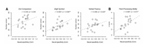
A central challenge in the study of healthy aging is to identify factors that distinguish those who age gracefully from those who do not, because doing so could shed light on the underlying causes of age-related declines and how best to address them. We recently discovered that neural distinctiveness might be an important factor. Fluid processing ability is known to decline with age and we showed that individual differences in neural distinctiveness predict performance on a range of fluid processing tasks in older adults (Park et al., 2010![]() ). In fact, neural distinctiveness accounted for 30% of the variance in behavioral performance, despite the fact that neural distinctiveness was measured in visual cortex using simple visual tasks while the fluid processing tasks required far more general types of cognitive processing:
). In fact, neural distinctiveness accounted for 30% of the variance in behavioral performance, despite the fact that neural distinctiveness was measured in visual cortex using simple visual tasks while the fluid processing tasks required far more general types of cognitive processing:
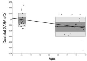
Another one of our main research questions is what underlies this age-related decline in neural distinctiveness? Research in animals has found a link between the brain’s major inhibitory neurotransmitter (gamma-aminobutyric acid or GABA for short) and the selectivity of neural receptive fields. We therefore decided to measure GABA levels in humans using magnetic resonance spectroscopy (MRS). We found that GABA levels in occipital cortex were significantly lower in older (59-87 years old) participants compared to younger participants (18-23 years old) (Simmonite et al., 2018![]() ) :
) :
We also observed a significant relationship between GABA levels and fluid processing ability suggesting that the decline in those abilities might be associated with the decline in GABA:

To further explore these topics, we recently began a multi-year research study: the Michigan Neural Distinctiveness (MiND) project. The study aims to investigate the scope of age-related neural dedifferentiation (i.e., does it occur in auditory and motor cortex like it does in visual cortex?), potential causes (age-related reductions in GABA levels), and behavioral consequences (age-related decline in cognitive abilities). We are using fMRI and MR spectroscopy to measure neural distinctiveness and GABA concentrations in the same brain region in older and younger adults (Gagnon et al., 2019![]() ). Additionally, we are collecting an extensive battery of behavioral measures in the same subjects, allowing us to observe individual differences in neurochemistry, neural representation, and behavioral performance, rather than focusing on group differences (young vs. old) alone.
). Additionally, we are collecting an extensive battery of behavioral measures in the same subjects, allowing us to observe individual differences in neurochemistry, neural representation, and behavioral performance, rather than focusing on group differences (young vs. old) alone.
A recent publication from the MiND study investigated whether neural dedifferentiation extends to auditory cortex and whether differences in GABA are associated with individual differences in neural distinctiveness. We observed that compared to younger adults, older adults exhibited less distinct activation patterns for music vs. speech stimuli and lower GABA levels in auditory cortex (Lalwani et al., 2018![]() ). Also, individual differences in auditory GABA levels predicted individual differences in neural distinctiveness auditory cortex in the older adults.
). Also, individual differences in auditory GABA levels predicted individual differences in neural distinctiveness auditory cortex in the older adults.
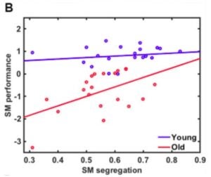
Researchers interested in aging have also observed age-related neural differentiation between neural networks observed at rest. Specifically, these networks are typically less segregated from each other in older vs. younger participants (another form of dedifferentiation). We therefore also investigated the relationship among network segregation, GABA, and sensorimotor function. We observed that older age is associated with reduced segregation and reduced GABA levels in sensorimotor cortex (Cassidy et al., 2019![]() ). Reduced network segregation and GABA levels, in turn, correlated with worse sensorimotor function among older adults :
). Reduced network segregation and GABA levels, in turn, correlated with worse sensorimotor function among older adults :
Furthermore, we found that network segregation mediated the link between GABA and sensorimotor performance:

These results provide a rare link from the brain’s neurochemistry through its functional connectivity all the way to observable behavior.
Completed projects:
- The role of GABA in neural dedifferentiation in the visual cortex (led by Jordan Chamberlain). Learn more
- The relationship between sensorimotor network segregation and GABA (led by Kaitlin Cassady) Learn more
- The relationship between network segregation and neural distinctiveness in sensorimotor cortex (led by Kaitlin Cassady) Learn more
- The relationship between auditory GABA and neural distinctiveness (led by Pia Lalwani) Learn more
- Age-related changes in white matter (led by Shannon Kelley) Learn more
Current projects in the lab are exploring a number of related topics:
- The relationship between GABA and neural variability (led by Pia Lalwani)
- The relationship between fMRI and EEG measures of neural dedifferentiation (led by Ryan Rich and Pia Lalwani) Learn more
- Age-related changes in GABA and other metabolites across the brain (led by Molly Simmonite) Learn more
- Whether neural dedifferentiation in one brain region is associated with neural dedifferentiation in other regions (suggesting a common cause) (led by Molly Simmonite)
- The relationship between MRS measures of GABA and inhibitory function measured using transcranial magnetic stimulation (led by Dalia Khammash) Learn more
Future projects:
-
Investigating age-related neural dedifferentiation longitudinally and in Alzheimer’s pathology: A growing body of research has found that neural representations are less distinctive in older relative to younger adults, a phenomenon known as age-related neural dedifferentiation. Neural dedifferentiation has been associated with many of the behavioral impairments typically observed in healthy aging, and evidence collected by our group during the original funding period has found a strong relationship between neural dedifferentiation and gamma-aminobutyric acid (GABA), the brain’s major inhibitory neurotransmitter. Specifically, we found that GABA levels are lower in older vs. younger adults and that older adults with less GABA exhibit greater dedifferentiation. We propose to take this line of research in two new directions. (1) We will conduct a longitudinal study and explore age-related trajectories of change in GABA, neural distinctiveness, and behavior as well as directional relationships among these measures (e.g., Do GABA levels at age 65 predict subsequent declines in distinctiveness? Does distinctiveness at 65 predict subsequent behavioral declines?). (2) We will examine how dedifferentiation is related to Alzheimer’s pathology by collecting the same measures in patients in whom amyloid beta and tau burden have already been measured.